PEMPix is the American Academy of Pediatrics Section on Emergency Medicine’s annual visual diagnosis competition. This year, in addition to the 10 finalists I will be presenting at the National Conference and Exhibition I will be sharing four cases online in advance of the conference. This is the first of the four cases.

This case was submitted by…

A previously healthy three year-old female presents from pediatrician’s office for evaluation of two months of weight loss. Specifically she has had a twelve pound weight loss over the last two months with decreased activity, generalized pain, rash, and bruising for the last week. She has also had daily subjective fevers for the about the last week. Her mother thinks the child is having joint pain, but the cannot localize it. She has also been to other emergency rooms in the past two months, but was told it was likely viral and to follow up both the with pediatrician.

Prior to this illness, the child was happy and playful with a good appetite. Now she only eats a few bites of food at a meal, and she seems to have trouble swallowing liquids – which she has been choking on over the last few days. She is mostly sedentary now, and becomes fatigued very easily. She also had a couple episodes of ”black” stools two weeks ago, and two days prior to ED arrival developed mild dry cough and rhinorrhea. There is no increased work of breathing, no chest pain, and no bleeding from nose or mouth. There is also no nausea, vomiting, or diarrhea. She is urinating less however. There are no sick contacts, recent travel, or tuberculosis exposures. The family is from Cuba, but patient was born in the United States.

Physical Examination

Vital signs BP 130/86 | Pulse 148 | Temp 100.1 °F | Resp 24 | Wt 14.8 kg (31.85%tile)  | SpO2 96% 

Constitutional Cachectic, tired-appearing, sitting in mother’s lap, not wanting to be examined

HEENT Normocephalic, normal tympanic membranes, rhinorrhea, moist mucus membranes, PERRLA, conjunctiva normal

Heart RRR, no murmurs, good perfusion, pulses strong

Pulmonary effort normal, no wheezes, no rhonchi

Abdomen soft, nontender, no distention, liver 1cm below costal margin

Musculoskeletal No point tenderness, no swelling, no deformities, no vertebral tenderness. Shuffling gait. Pain with extension/flexion of bilateral wrists and ankles. Bilateral knee swelling with erythema. Uncooperative on focal strength testing, movements of upper extremities are slow, but he is able to sit up and get off couch without assistance, does roll over from stomach to back

Lymphatic No cervical, axillary, inguinal lymphadenopathy

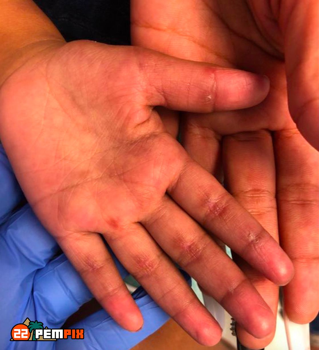
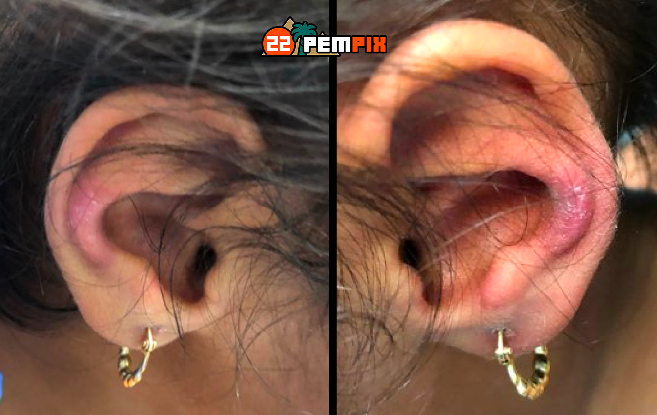
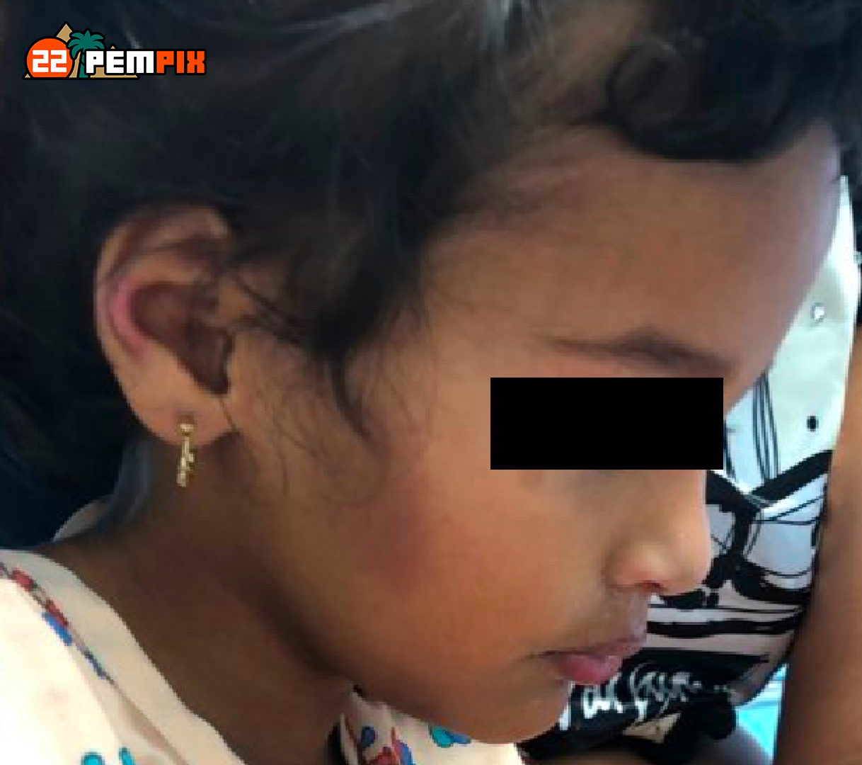
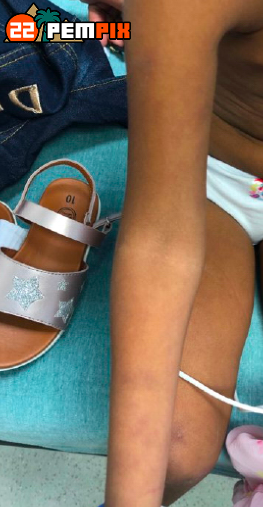
Skin ecchymosis over bilateral knees, right shin, right cheek, bilateral wrists. Faint purple periorbital discoloration with petechiae on upper left eyelid. Faint erythematous rash over bilateral elbows, bilateral MCPs, bilateral knees, lower abdomen. Dry, cracked areas on palmar surface of hands bilaterally at PIPs. Dry, erythematous rash over bilateral ears

Neurological no focal deficits

Labs

Abnormal findings in bold

  • WBC 8.86 x 10⁹/L – Hgb 11.4 g/L – Hct 34.6 – Platlets 236 x 109/L
  • Na 138, K 4, Cl 105, CO2 25, BUN 5, Cr 0.15, Gluc 92, Mg 1.9, Ca 9
  • ALT 180, AST 459, GGT 150, Bili 0.3, Lipase 95, Prealbumin 8.6
  • LDH 1255, Uric Acid 2.6, CRP <0.5, CK 94, ESR 60
  • INR 1.0, PT 14, PTT 40
  • TSH 1.063, T4 20.6
  • Urinalysis – Trace ketones, WBC 5-10, Bacteria trace, nitrite negative, protein negative

Imaging

  • Abdominal ultrasound showed hepatosplenomegaly, and no additional abnormalities
  • Chest X-Ray was normal

A. Child Neglect

B. Dermatomyositis

C. Celiac disease

D. Hyperthyroidism

E. Systemic lupus erythematosus

B. Dermatomyositis

The diagnosis was most supported by the heliotrope rash on her knuckles, as well as rash over extensor surfaces, proximal weakness, evidence of vasculopathy with GI involvement (dysphagia, possible melena), bruising, elevated LDH and liver transaminases.

Dermatomyositis is an extremely rare disease (3 cases per 1,000,000) that is due to inflammation of blood vessels and striated skeletal muscle It is more common in females. Diagnostic criteria include:

  • Symmetric proximal muscle limb weakness
  • Necrosis of type I and II muscle fibers on biopsy
  • Elevated creatine phosphokinase and aldolase
  • Abnormal electromyography
  • Heliotrope rash

The disease has 4 phases: prodromal (aches and pains), progressive muscle and skin inflammation with weakness and rash, persistent active muscle damage, and an indolent phase with development of contractures and calcinosis. You will see a wide variety of enzymes elevated: CK, aldolase, LDH, and Transaminases. Complications include GI hemorrhage, respiratory failure, velopalatine weakness, pneumothorax, and carditis.

The goals of treatment are to control muscle inflammation. Often this means starting with pulse IV methylprednisolone, then switching to oral prednisone. If no improvement with steroids is seen, or in severe disease, patients will receive IVIG, methotrexate, and/or cyclosporine.

Why wasn’t it?

Child Neglect

Mom was appropriately seeking care and the child was followed closely by pediatrician.

Celiac disease

No diarrhea or abdominal pain and the rash with celiac disease normally found on elbows, knees and buttocks – not the fingers and ears, and it itches intensely.

Hyperthyroidism

Normal TSH and elevated T4 concerning for hyperthyroidism, but rash and proximal weakness favors dermatomyositis

Systemic Lupus Erythematosus

No malar rash – and the symptoms pointed elsewhere.

Patient Follow-Up

She was treated with steroids, methotrexate, and IVIG. Hospitalization was complicated by the development of severe dysphagia necessitating peripheral parenteral nutrition. She later developed interstitial lung disease, respiratory failure, and sadly passed away approximately 3 weeks after ED presentation.

References

kLundberg IE, Tjärnlund A, Bottai M, Werth VP, Pilkington C, de Visser M, Alfredsson L, Amato AA, Barohn RJ, Liang MH, Singh JA, Aggarwal R, Arnardottir S, Chinoy H, Cooper RG, Dankó K, Dimachkie MM, Feldman BM, Garcia-De La Torre I, Gordon P, Hayashi T, Katz JD, Kohsaka H, Lachenbruch PA, Lang BA, Li Y, Oddis CV, Olesinska M, Reed AM, Rutkowska-Sak L, Sanner H, Selva-O’Callaghan A, Song YW, Vencovsky J, Ytterberg SR, Miller FW, Rider LG; International Myositis Classification Criteria Project Consortium, the Euromyositis Register, and the Juvenile Dermatomyositis Cohort Biomarker Study and Repository (UK and Ireland). 2017 European League Against Rheumatism/American College of Rheumatology Classification Criteria for Adult and Juvenile Idiopathic Inflammatory Myopathies and Their Major Subgroups. Arthritis Rheumatol. 2017 Dec;69(12):2271-2282. doi: 10.1002/art.40320. Epub 2017 Oct 27. Erratum in: Arthritis Rheumatol. 2018 Sep;70(9):1532. PMID: 29106061; PMCID: PMC5846474.
 
Lundberg IE, Tjärnlund A, Bottai M, Werth VP, Pilkington C, Visser M, Alfredsson L, Amato AA, Barohn RJ, Liang MH, Singh JA, Aggarwal R, Arnardottir S, Chinoy H, Cooper RG, Dankó K, Dimachkie MM, Feldman BM, Torre IG, Gordon P, Hayashi T, Katz JD, Kohsaka H, Lachenbruch PA, Lang BA, Li Y, Oddis CV, Olesinska M, Reed AM, Rutkowska-Sak L, Sanner H, Selva-O’Callaghan A, Song YW, Vencovsky J, Ytterberg SR, Miller FW, Rider LG; International Myositis Classification Criteria Project consortium, The Euromyositis register and The Juvenile Dermatomyositis Cohort Biomarker Study and Repository (JDRG) (UK and Ireland). 2017 European League Against Rheumatism/American College of Rheumatology classification criteria for adult and juvenile idiopathic inflammatory myopathies and their major subgroups. Ann Rheum Dis. 2017 Dec;76(12):1955-1964. doi: 10.1136/annrheumdis-2017-211468. Epub 2017 Oct 27. Erratum in: Ann Rheum Dis. 2018 Sep;77(9):e64. PMID: 29079590; PMCID: PMC5736307.