PEMPix is the American Academy of Pediatrics Section on Emergency Medicine’s annual visual diagnosis competition. This year, in addition to the 10 finalists I will be presenting at the National Conference and Exhibition I will be sharing four cases online in advance of the conference. This is the third of the four cases.


This case was submitted by…


Dr. Roach was assisted on presenting this case by Daisy Ciener, MD, MS, a Pediatric Emergency Medicine Attending Vanderbilt.


A 12-year-old male with a history of developmental delay and a prior nasal tumor status post resection, was transferred from an outside hospital for abdominal pain, bloody and bilious emesis, and diarrhea for 3 days. On the morning prior to presentation, the patient was seen by his pediatrician he was given ondansetron and told to go to the ED if his symptoms worsened. That night, his symptoms worsened, and Mom took him to the community ED where he had diffuse, but non-specific abdominal tenderness. He received IV ondansetron and a 20ml/kg normal saline bolus, and labs were notable for:


  • Platelets 433 x103 mcL
  • CRP 10.7 mg/L
  • BMP, urinalysis, strep, and viral testing were negative


No imaging studies were obtained at this initial visit, and he returned to this facility on the next day (the day of transfer to our ED) with worsening abdominal distention, pain, and vomiting. His vitals were initially normal, but he quickly deteriorated with systolic blood pressure reaching the 70s. On exam he was ill appearing with abdominal distention, guarding, and rebound tenderness. Notable labs included:


  • Na 147 mmol/L
  • Creatinine 1.86 mg/dL
  • Glucose 196 mg/dL
  • Anion Gap 24
  • White Blood Cell Count 13.4 x10³ mcL
  • Platelets 424 x103 mcL
  • C-Reactive Protein 76.7 mg/L
  • Lactate 12.4 mmol/L


He was given 1L NS, pipericillin / tazobactam, morphine , and ondansetron. A CT of the abdomen and pelvis was obtained prior to transfer:


A. Gastric Bezoar

B. Duodenal Atresia

C. Malrotation with Midgut Volvulus

D. Gastric Volvulus

E. Abdominal Rhabdomyosarcoma


D. Gastric Volvulus

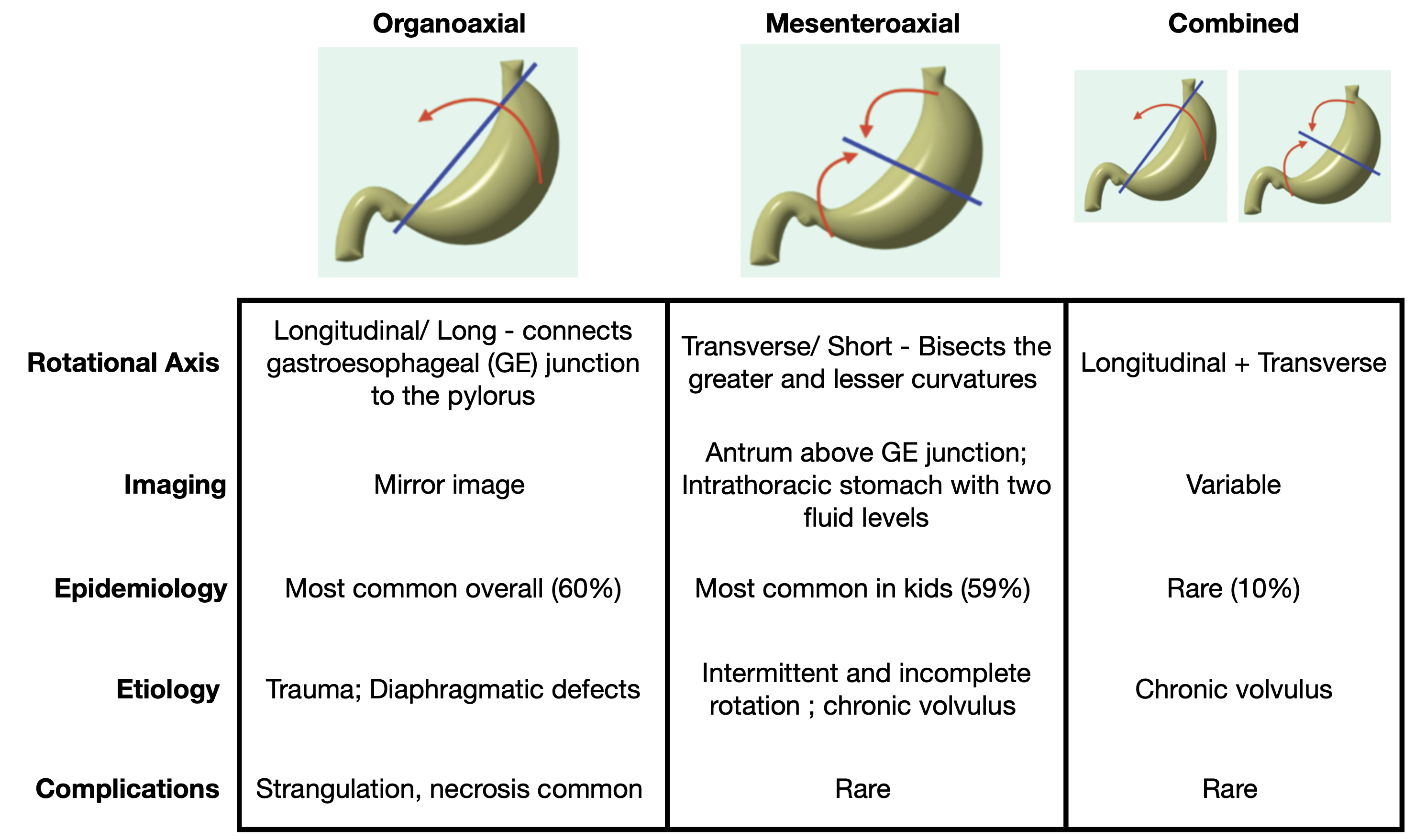
Gastric volvulus is defined as abnormal rotation of the stomach >180 degrees. Normally, the stomach is fixed in position by 4 ligaments (gastro-colic, gastro-hepatic, gastro-phrenic, and gastro-splenic). Gastric volvulus presents with pain, vomiting, and patients become rapidly sicker due to abdominal distention, acidosis, and gut necrosis. 80% of cases occur in adults, and the mortality is as high as 50%. It is classified in the following manner:

  • According to type = organoaxial, mesenteroaxial, combined 
  • According to etiology = primary or secondary
    • Primary = 2/3 cases, Idiopathic; Due to laxity or absence of gastric ligaments
    • Secondary = 1/3 cases; due to anatomic or functional abnormalities that result in abnormal mobility of stomach: Diaphragmatic defects, eventration of the diaphragm, Paraesophageal hernia, Abnormal attachments / adhesions / bands, Pyloric stenosis, Congenital asplenia, Bowel malformations, colonic distention
  • According to presentation = acute or chronic 

Images from: Al-Salem, A.H. (2020). Atlas of Pediatric Surgery. Springer, Cham.

Why wasn’t it?

Gastric Bezoar

A foreign body in the stomach resulting from the accumulation of ingested material; classified according to their composition (vegetables, hairs, medications, other); insidious onset. It is generally found incidentally on imaging. 

Duodenal atresia

Most common cause of congenital duodenal obstruction, and presents with bilious emesis. It is typically associated with other congenital anomalies.  

Malrotation with midgut volvulus

Occurs due to arrest of normal rotation of the embryonic gut; bowel twists around the superior mesenteric artery; classically presents in infancy as bilious emesis however 22% of older children with malrotation present with volvulus.

Abdominal Rhabdomyosarcoma

Most common soft tissue sarcoma in children; associated with Li-Fraumeni Syndrome, NF1, BWS, Dicer1 Syndrome, and Noonan Syndrome; can present with abdominal pain/ distention and vomiting.

Patient Follow-Up

The patient went straight to the OR with pediatric surgery for total gastrectomy. Unfortunately he succumbed to septic shock and died some five weeks later. 

References

Al-Salem, A.H. (2020). Gastric Volvulus. In: Atlas of Pediatric Surgery. Springer, Cham. https://doi-org.proxy.library.vanderbilt.edu/10.1007/978-3-030-29211-9_46

Aslam, Shehroz MD; Alani, Mustafa MD; Ansari, Zaid MD; Moussa, Jacob MD; Srinivasan, Indu M; Chuang, Keng-Yu MD3 S3156. Gastric Volvulus: An Underrated Complication of Hiatal Hernia, The American Journal of Gastroenterology: October 2021 – Volume 116 – Issue – p S1301 doi: 10.14309/01.ajg.0000786156.67258.10

Arun, Ganesh DO; Rey, Emily DO; Varughese, Dimple DO; Reddy, Dayakar MD; Patel, Ami DO S3712 Gastric Volvulus: A Difficult Hernia to Swallow, The American Journal of Gastroenterology: October 2021 – Volume 116 – Issue – p S1515-S1516 doi: 10.14309/01.ajg.0000789196.56848.fb

Brinkley MF, Tracy ET, Maxfield CM. Congenital duodenal obstruction: causes and imaging approach. Pediatric Radiology. 2016;46(8):1084-1095. doi:10.1007/s00247-016-3603-1

Cianci MC, Coletta R, Morabito A. Let’s (Not) Twist Again: Laparoscopic-Assisted Percutaneous Endoscopic Gastrostomy in Paediatric Gastric Volvulus. Digestive diseases and sciences. 2021;66(8):2533-2536. doi:10.1007/s10620-021-07071-6

Güven B, Beger B, Uslu E, Düz E. Gastric volvulus: a rare cause of gastric outlet obstruction in pediatric age. The European research journal. 2018;5(3):566-568. doi:10.18621/eurj.407805

Jones, J., Vadera, S. Gastric volvulus. Reference article, Radiopaedia.org. (accessed on 08 Jun 2022) https://doi.org/10.53347/rID-6170

Porcaro F, Mattioli G, Romano C. Pediatric Gastric Volvulus: Diagnostic and Clinical Approach. Case reports in gastroenterology. 2013;7(1):63-68. doi:10.1159/000348758