

It’s not the heart way more often than it is the heart when it comes to pediatric chest pain in the Emergency Department. The purpose of this post is to highlight factors in the history and physical examination that should make you think that it could actually be the heart. Note that some of this information is also applicable for children with syncope, and for completeness sake appears in that post as well.
This post specifically focuses on kids that are well appearing, and can help you make the decision on whether or not to refer a child to pediatric Cardiology versus follow up with their Primary Care Doctor.
When a child presents with chest pain you should be worried if…
The pain only occurs with exertion
Specifically, you should be worried if the pain occurs at peak exertion – not low level exercise, and does not occur at rest. So, sprinting and chest pain is more worrisome than pain that occurs with light jogging. Worrisome pain that occurs on peak exertion should also not be reproducible to palpation.
They had exertional syncope
Exertional syncope and chest pain are needless to say a worrisome combo, the classic examples of which would be hypertrophic and dilated cardiomyopathy, and left ventricular outflow tract obstruction. Patients will have murmurs, and oftentimes a positive family history of cardiomyopathy. Children may also have a history of declining exercise tolerance as well.
The pain is positional
Specifically, you should be concerned if retrosternal pain is worse when the patient is supine, sometimes radiating to the left shoulder, and is associated with fever. this combo should make you think of pericarditis. You don’t have to call Cards on every child with pericarditis – but if you’re not sure of your diagnosis, or you’re worried that they could have associated myocarditis as well by all means then get them on the phone.
Note: If they really do look like they are having a heart attack – with anginal pain that is crushing, substernal, and radiating down the left arm or up into the jaw – associated with vomiting, diaphoresis, altered mental status, or dyspnea – then you should manage appropriately. Again, this post is about kids who are appropriate for discharge home, and you are trying to decide whether to refer to Pediatric Cardiology or not.
Past Medical History
Obviously if the patient has a history of congenital heart disease that required a major intervention like surgery they should follow up with a cardiologist. This is a brief list of some other conditions that should prompt referral to Cardiology for follow up of chest pain.
Acquired heart disease or known predisposing condition
In a child or adolescent with chest pain you should always ask about history of Kawasaki Disease. As high as 1 in 4 patients with Kawasaki you were not treated will have cardiac complications. Treatment with IVIg lowers the risk substantially.
If you see a patient with chest pain, and a past medical history of Marfan, Loeys-Dietz, type IV Ehlers-Danlos, or Turner syndromes know that they will need evaluation for aortic root problems (and coarctation in the case of Turner). Fortunately, where I work these patients are followed closely by cardiology.
Conditions that predispose to pericarditis include rheumatologic disease, cancer, recent cardiac surgery, mediastinal radiation, renal failure, and infections such as tuberculosis and HIV.
Hypercoagulable state and thrombophilia
There are several of note – but even if the child looks well Cards should probably see someone with a history of iInherited hypercoagulable conditions including factor V Leyden, protein C or protein S deficiency, elevated homocysteine, dysfibrogeniemia and more.
At this point I think you know the stuff that makes you at risk for blood clots. Some of which include;
- Immobility from recent surgery or medical problem
- Oral contraceptive use
- Central line
- Solid tumor
- Obesity
Family History
Cardiomyopathy
Many non-medical folks will not know what this is. Cardiomyopathy is broadly defined, and occurs in a number of settings; post viral, in pregnancy, after an acute cardiac event and more. Furthermore, there are different subtypes, dilated, restrictive. I therefore recommend the following lay definition that will at least get across the gist of what Cardiomyopathy is;
“A disease of the heart muscle that makes it harder for that heart to pump blood to the rest of your body.”
MayoClinic.org
Sudden death in someone less than 50 years old
This one can be hard to nail down in the history as well. It can also dredge up difficult memories. Perhaps someone died of cardiac arrest following a drug overdose for instance. I would recommend asking specifically if a first degree relative died before they were 50, and the case was known to be due to the heart. For instance, a heart attack.
A known arrhythmia like long QT syndrome or Brugada syndrome
This is another one that people won’t know about unless they are in medicine, or have a family member with it. Again, describing the problem in lay terms, about an abnormal heart rhythm that is inherited or medication induced that can lead to syncope or even sudden death.
A family member under 50 has a pacemaker or defibrillator
It is relatively uncommon to get a pacemaker or implantable defibrillator when you are under 50. Even if they don’t know precisely why, it should at least make you curious about what kind of heart condition a family member has if they have a heart fixing computer embedded in their chest wall connected to their heart. Per Healey et al, the average age for pacer implantation is 75 years of age, with sick sinus syndrome and AV block being among the most common reasons. Only 6% of patients with a pacemaker are under 50.
A known familial coronary anomaly
I haven’t personally seen this in practice but there are rare cases of inherited coronary artery anomalies. Per Laureti et al.”the most serious problem is the predisposition for sudden cardiac death when the coronary artery follows an interarterial course between the aorta and the pulmonary artery.”
Specific EKG findings
QTc interval >470 msec
Remember, this is one that we calculate manually, using an accepted tool or equation. The one that many of us were taught is:
QTc = QT interval ÷ √RR interval [in sec]
Long QT syndrome can lead to syncope and sudden cardiac death. So if the calculated measurement (not the one the EKG spit out) is longer than 470 msec refer the patient to Cardiology.
Pre-excitation
Patients with pre-excitation have an accessory pathway that bypasses the AV node. Wolff-Parkinson-White syndrome is the existence of pre-excitation on EKG and an episode of tachycardia. Per an excellent post on Life in the Fast Lane the main EKG features of pre-excitation are:
- PR interval <120ms
- “Delta wave” slurring slow rise of initial portion of the QRS
- QRS prolongation >110ms
- ST Segment and T wave discordant changes – i.e. in the opposite direction to the major component of the QRS complex
- Pseudo-infarction pattern can be seen in up to 70% of patients – due to negatively deflected delta waves in the inferior / anterior leads (“pseudo-Q waves”), or as a prominent R wave in V1-3 (mimicking posterior infarction)
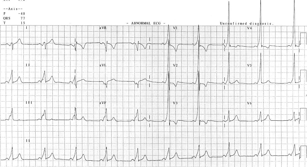
Brugada pattern
Brugada is a pseudo-right bundle branch block (RBBB) and persistent ST segment elevation in leads V1 to V3. You’ll see ST segment elevation >2mm in >1 of V1-V3 followed by a negative T wave. It is a genetic sodium channel mutation. A sample of the waveform is seen below. The mean age of sudden death is 41. It has a much higher prevalence in Southeast Asia.

Abnormal voltage and intervals
You need to have access to age and gender-based norms and make sure you calculate all of the peaks an intervals. This is part of doing the work to make sure a patient is safe or if the need to be referred to Cardiology. There are some simple “hacks” you can remember though. Abnormal findings for ventricular hypertrophy, adapted from Evans et al, and borrowed from Ped EM Morsels include:
- Abnormal Left Ventricular Large Voltage (“LVH”)
- Use only V6 (the left most precordial lead)
- If R wave of V6 intersects with baseline of V5, then that is abnormal.
- Abnormal Right Ventricular Large Voltage (“RVH”)
- Use only V1 (the right most precordial lead)
- Upright T wave in V1?
- During 1st week of life, T wave can be upright in V1.
- After 1st week of life, upright T wave in V1 is abnormal in children until adolescence.
- With RSR’ is present, if R’ is taller than R wave, then this is abnormal.
- A pure R wave in V1 in a child > 6 months of age is abnormal.
You should also refer for First degree AV block with a PR interval >250 msec.
Pathological ST segment changes
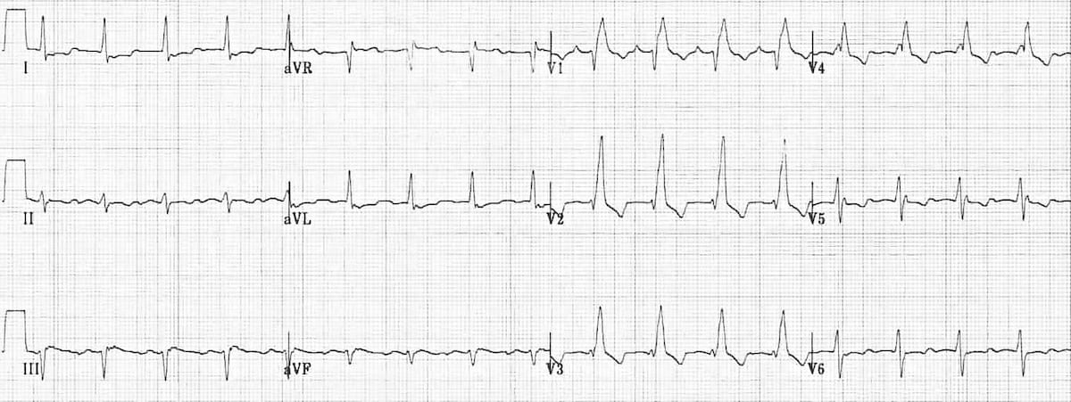
The main point that I want to make here is that many healthy teens will have a subtle upsloping of the ST segment immediately after the QRS complex. This is sometimes called “J-point elevation” or early re-polarization, and is seen in healthy young hearts. ST elevation in grown ups makes one think of myocardial infarct. Benign early depolarization can be seen in 1 of 10 patients with chest pain presenting to the ED, so how do you tell the difference? First, get good at looking at EKGs. Here are some examples of early depolarization, again courtesy of Life in the Fast Lane:


It is also common to see “notching” of the J-point in benign early depolarization. This gives the ST-segment a “fish hook” appearance as seen in the embedded image. This notching is generally best seen in lead V4.
Note that early depolarization can be more prominent with slower heart rates. There is also a validated calculator that will help you differentiate early depolarization from STEMI. It is most useful in patients with a suspicious history for MI, but a non-diagnostic EKG. It involves calculating/measuring the following 4 things and using the following formula:
Subtle Anterior STEMI 4-Variable Calculation = 0.052 x (Bazett-corrected QT interval, ms) - 0.151 x (QRS amplitude in lead V2, mm) - 0.268 x (R wave amplitude in lead V4, mm) + 1.062 x (ST segment elevation 60 ms after the J point in lead V3, mm)
Or you could just use the calculator found at MDCalc. Your choice…
Abnormal T-wave inversion
You should commit to memory, or at least find a way to remember when the t waves flip around in life. These are some general rules:
- First week of life: Upright T waves in precordial leads
- After the first week u until age ~8 years: T waves inverted in V1-3
- After 8-years: T waves become upright in V1-3
- Some patients can have this juvenile pattern with upright T waves in V1-3 that persists past adolescence into the early 20s
Bundle branch block
Right bundle branch block
Criteria from Life in the Fast Lane
- Broad QRS > 120 ms
- RSR’ pattern in V1-3 (‘M-shaped’ QRS complex)
- Wide, slurred S wave in the lateral leads (I, aVL, V5-6)
Left bundle branch block
Criteria from Life in the Fast Lane
- QRS duration of > 120 ms
- Dominant S wave in V1
- Broad monophasic R wave in lateral leads (I, aVL, V5-V6)
- Absence of Q waves in lateral leads (I, V5-V6; small Q waves are still allowed in aVL)
- Prolonged R wave peak time > 60ms in left precordial leads (V5-6)
Examination findings
Some specific examples include:
- A systolic ejection murmur and/or an ejection click which are heard in aortic stenosis
- A difference in pulse quality in upper and lower extremities (upper > lower), and a difference in upper and lower extremity systolic blood pressures arm 20 mmHg or more greater than leg, can be seen in coarctation of the aorta
- A murmur that is heard in hypertrophic cardiomyopathy is one that decreases in intensity with increased venous return to the heart (during a Valsalva or squatting)
- Rales on lung exam (not really the heart exam)
- A friction rub or gallop
- A LOUD S2
- Hepatosplenomegaly (unless the patient concurrently has mono and faints) can be indicative of heart failure
- Rales on lung exam are also a sign of heart failure
References
Burns. Left Bundle Branch Block. Life in the Fast Lane. https://litfl.com/right-bundle-branch-block-lbbb-ecg-library/ April 27, 2019. Accessed February 8, 2020.
Burns. Right Bundle Branch Block. Life in the Fast Lane. https://litfl.com/right-bundle-branch-block-rbbb-ecg-library/ March 16, 2019. Accessed February 8, 2020.
Evans WN, Acherman RJ, Mayman GA, Rollins RC, Kip KT. Simplified pediatric electrocardiogram interpretation. Clin Pediatr (Phila). 2010 Apr;49(4):363-72. PMID: 20118092
Fox S. Pediatric ECG. Pediatric EM Morsels. https://pedemmorsels.com/pediatric-ecg/. September 8, 2017. Accessed February 7, 2020.
Friedman KG, Alexander ME. Chest pain and syncope in children: a practical approach to the diagnosis of cardiac disease. J Pediatr. 2013 Sep;163(3):896-901.e3. Epub 2013 Jun 12.
Laureti et al. Anomalous coronary arteries: A familial clustering. Clin Cardiology, 2005.
O’Connor M, McDaniel N, Brady WJ. The pediatric electrocardiogram. Part I: Age-related interpretation. Am J Emerg Med. 2008 Feb;26(2):221-8. PMID: 18272106


